
Electron Tomography
|
Electron tomography uses information much like that obtained in hospitals as images from CAT scans and MRIs to provide a full three-dimensional picture inside microscopic objects. But while CAT scans use x rays to form the images and MRIs use magnetic properties of atoms to form the images, here we use electrons that are transmitted through the sample to form images with sub-nanometer spatial resolution. This approach offers the unique ability to sort out material types in very small sample sizes, as well as cleverly using the two dimensional data to reconstruct amazingly detailed three dimensional pictures of complex structures with dimensions in the nanoscale regime. |
|||
|
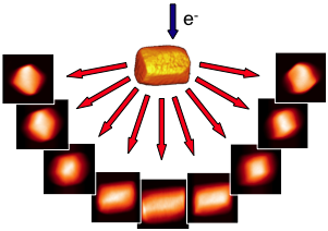
Multiple projections of the sample are recorded at many different orientations |
The projected data is recombined using backprojection algorithms to reconstruct the original object |
||

 |
 |
 |
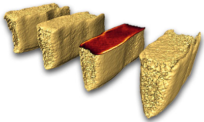
Three-dimensional imaging of nanovoids, roughness and critical dimensions in electrical interconnects using electron tomography.These projects are performed in collaboration with our partners at AMD, Applied Materials, IBM, Intel and Novellus. The work is funded by the Semiconductor Research Corporation and its member companies and NYSTAR, with facilities support from the National Science Foundation. |
Three-dimensional reconstruction of silicon nanoparticles by plasmon imagingWork supported by theNational Science Foundation through the Cornell Center for Nanoscale Systems with facilities support from the Cornell Center for Materials Research. |
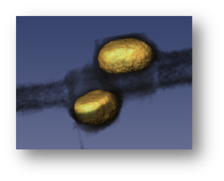
Three-dimensional studies of how and when metal contacts can deform carbon nanotubesWork supported by the National Science Foundation through the Cornell Center for Materials Research. |